Symptoms

Τενοντίτιδα ώμου

Shoulder tendonitis | Diagnosis & treatment

Shoulder tendonitis is a common condition characterized by inflammation of the rotator cuff or biceps tendon, often resulting from repetitive overhead movements or structural impact. This article explores the causes, symptoms, diagnostic methods, and treatment options, while discussing its relationship to impingement syndrome and rotator cuff tears.

What is shoulder tendonitis?

Η τενοντίτιδα ώμου είναι η φλεγμονή των τενόντων που συνδέουν τους μύες του στροφικού πετάλου (υπερακάνθιος, υποακάνθιος, έλασσων στρογγύλος και υποπλάτιος) με την κεφαλή του βραχιονίου. Αυτοί οι τένοντες σταθεροποιούν την άρθρωση του ώμου και διευκολύνουν την κίνηση. Όταν παρουσιάζουν φλεγμονή, οι τένοντες παχύνονται και μπορεί να υφίστανται παγίδευση κάτω από το ακρώμιο (μια οστική προεξοχή της ωμοπλάτης), οδηγώντας σε πόνο και περιορισμένη κινητικότητα. Αυτή η τελευταία κατάσταση είναι γνωστή και ως impingement syndrome ώμου, ή σύνδρομο υπακρωμιακής προστριβής. Πιο συχνή είναι η τενοντίτιδα υπερακανθίου.
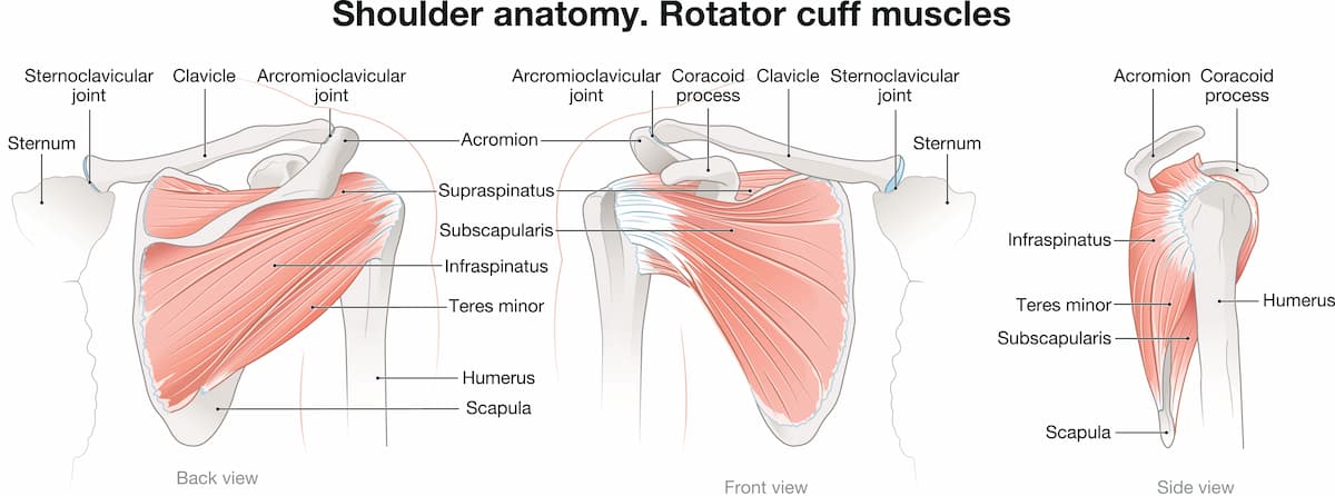
Ανατομία ώμου
Normal shoulder anatomy

Causes & risk factors

Causes of shoulder tendonitis – shoulder impingement syndrome include:

Επαναλαμβανόμενες κινήσεις πάνω από το κεφάλι: Συχνές σε αθλήματα όπως το μπέιζμπολ, το κολύμπι, το τένις και η άρση βαρών, καθώς και σε επαγγέλματα όπως οι ελαιοχρωματιστές ή οι ξυλουργοί.

Κακή στάση ή τεχνική: προκαλεί επιπλέον έργο και επιβαρύνει τους τένοντες.

Age: Οι τένοντες χάνουν την ελαστικότητά τους με την ηλικία, κάνοντάς τους επιρρεπείς σε εκφυλισμό.

Οξείες κακώσεις: Πτώσεις ή ξαφνικές κινήσεις.

Οι παράγοντες κινδύνου περιλαμβάνουν:

  • Αθλήματα που σχετίζονται με επαναλαμβανόμενη κίνηση, όπως κολύμπι, τένις, ή volley
  • Εργασίες που προϋποθέτουν επαναλαμβανόμενη κίνηση πάνω από το κεφάλι
  • Οι δουλειές καθαριότητας του σπιτιού
  • Υποκείμενες παθήσεις που προκαλούν stress στον τένοντα, όπως αρθρίτιδα ή διαβήτης
  • Το ακατάλληλο ζέσταμα πριν από την προπόνηση

Αθλητής τένις με τενοντίτιδα ώμου

Symptoms

Τα συμπτώματα της τενοντίτιδας ώμου περιλαμβάνουν:

  • Πόνος στον ώμο κατά τη διάρκεια δραστηριοτήτων πάνω από το επίπεδο των ώμων (π.χ. φθάνοντας ένα ράφι ή ένα ντουλάπι, χτένισμα).
  • Νυχτερινός πόνος στον ώμο, που διαταράσσει την ποιότητα του ύπνου
  • Δυσκαμψία και αδυναμία του άκρου

Οι ρήξεις του στροφικού πετάλου (μερικές ή ολικές, συνήθως ρήξη υπερακανθίου) μπορεί να προκαλέσουν οξύ πόνο και σημαντική αδυναμία. Η ρήξη τένοντα ώμου συχνά αναπτύσσεται σαν εξέλιξη μετά από χρόνια τενοντίτιδα ή οξύ τραύμα.

Diagnosis

Dr Panagopoulos will take a full history and perform a detailed physical exam, including measuring range of motion and strength. An x-ray will be ordered to look for arthritis or calcific tendonitis. An ultrasound in the office will be performed to look for tears. An MRI may be ordered to further clarify your anatomy and the extent of the damage, like tear size or degree of muscle atrophy.

Non-surgical treatment

Conservative treatment of rotator cuff tears includes:

  • Rest
  • Activity modification
  • NSAIDS
  • Nonsteroidal anti-inflammatory drugs (NSAIDs)
  • Physiotherapy/home exercise program for strengthening and stretching exercises
  • Ένεση κορτιζόνης ή PRP

Conservative treatment will help the majority of patients improve their symptoms and overcome shoulder pain, returning to daily activities. If symptoms do not subside, the next option is surgical repair with shoulder arthroscopy.

Γυναίκα με τενοντίτιδα ώμου

Surgical treatment

Σε περίπτωση που η συντηρητική θεραπεία δεν προσφέρει τα επιθυμητά αποτελέσματα, το επόμενο βήμα περιλαμβάνει την χειρουργική θεραπεία, με αρθροσκόπηση ώμου. Η αρθροσκόπηση ώμου γίνεται με 3 μικρές οπές 4 χιλιοστών γύρω από τον ώμο, μέσω των οποίων ο χειρουργός εισάγει μια κάμερα και διάφορα εργαλεία. 

This is a day procedure. It is performed under general +/- regional anesthetic. You can go home the same day if you wish, just a few hours after the procedure.

Κατά την αρθροσκόπηση ώμου, πραγματοποιείται χειρουργικός καθαρισμός της φλεγμονής του ώμου και λείανση του ακρωμίου προκειμένου να αυξηθεί ο διαθέσιμος χώρος για τους τένοντες του ώμου και να αποφευχθεί η τριβή. Στην συνέχεια, ο κ. Παναγόπουλος θα επιδιορθώσει τυχόν ρήξεις τένοντα με τη βοήθεια ειδικών ραμμάτων (άγκυρες). 

Η αρθροσκόπηση ώμου προσφέρει άριστα αποτελέσματα με εξαιρετικά χαμηλό ποσοστό επιπλοκών και προσιτό κόστος. Ο ασθενής εξέρχεται από το νοσοκομείο την ίδια μέρα, λίγες ώρες μετά του χειρουργείο. Το άκρο παραμένει σε ανάρτηση για κάποιο διάστημα, ανάλογα με την υποκείμενη παθολογία που αντιμετωπίστηκε. Ο ασθενής αρχίζει στη συνέχεια φυσικοθεραπεία. 

Η φυσικοθεραπεία είναι εξαιρετικά σημαντική κατά τη διάρκεια της αποκατάστασης μετά από αρθροσκόπηση ώμου. Ξεκινάει με ήπιες ασκήσεις άμεσα μετεγχειρητικά. Στη συνέχεια ακολουθούν ασκήσεις για την αποκατάσταση του εύρους κίνησης & ενδυνάμωση. Ο χρόνος αποκατάστασης εξαρτάται σε μεγάλο βαθμό από την υποκείμενη παθολογία.

Αν έχετε πόνο στον ώμο, είναι σημαντικό να επισκεφτείτε έναν εξειδικευμένο χειρουργό ώμου. Ο κ. Παναγόπουλος θα συζητήσει μαζί σας όλες τις εναλλακτικές θεραπείες κατά τη διάρκεια της επίσκεψής σας στο ιατρείο.

How effective are cortisone injections against shoulder tendonitis?

Οι ενέσεις κορτιζόνης είναι μια κοινή θεραπεία για την τενοντίτιδα ώμου, προσφέροντας σημαντικά βραχυπρόθεσμα οφέλη, προκαλώντας μείωση της φλεγμονής και του πόνου. 

Αποτελεσματικότητα των ενέσεων κορτιζόνης

1. Ανακούφιση του πόνου και μείωση της φλεγμονής: Οι ενέσεις κορτιζόνης είναι αποτελεσματικές στην ανακούφιση από τον πόνο και στη βελτίωση της λειτουργικότητας του ώμου, μειώνοντας τη φλεγμονή στην πληγείσα περιοχή. Μελέτες δείχνουν ότι μπορούν να προσφέρουν ανακούφιση για αρκετές εβδομάδες έως μήνες.

2. Σύγκριση με άλλες θεραπείες: Οι ενέσεις κορτιζόνης είναι γενικά πιο αποτελεσματικές από τα ΜΣΑΦ για βραχυπρόθεσμη ανακούφιση από τον πόνο στην τενοντίτιδα. Ωστόσο, τα μακροπρόθεσμα οφέλη τους είναι λιγότερο σαφή και μπορεί να μην είναι τόσο αποτελεσματικά όσο τα ΜΣΑΦ σε ορισμένες περιπτώσεις.

3. Δόση και διάρκεια: Υψηλότερες δόσεις κορτικοστεροειδών μπορεί να προσφέρουν καλύτερα αποτελέσματα από χαμηλότερες δόσεις, αν και αυτό απαιτεί προσεκτική εξέταση λόγω πιθανών παρενεργειών.

4. Συνδυαστική θεραπεία: Οι ενέσεις κορτιζόνης χρησιμοποιούνται συχνά σε συνδυασμό με ασκήσεις φυσικοθεραπείας και αποκατάστασης για την ενίσχυση της ανάρρωσης επιτρέποντας στους ασθενείς να συμμετέχουν σε ασκήσεις χωρίς πόνο.

Πιθανοί κίνδυνοι και επιπλοκές

1. Αποδυνάμωση τενόντων: Οι ενέσεις κορτιζόνης μπορεί να αποδυναμώσουν τους τένοντες με την πάροδο του χρόνου, κάτι που προκαλεί ανησυχία για ασθενείς που μπορεί τελικά να χρειαστούν χειρουργική επέμβαση αποκατάστασης στροφικού πετάλου.

2. Χειρουργικές επιπτώσεις: Πρόσφατες μελέτες υποδεικνύουν ότι οι ενέσεις κορτιζόνης πριν από τη χειρουργική επέμβαση του στροφικού πετάλου μπορεί να επηρεάσουν αρνητικά την επούλωση της χειρουργικής αποκατάστασης.

3. Παρενέργειες: Αν και γενικά είναι καλά ανεκτές, οι ενέσεις κορτιζόνης μπορεί να προκαλέσουν τοπικές παρενέργειες, όπως λέπτυνση ή αποχρωματισμό του δέρματος. Οι συστηματικές επιδράσεις, όπως τα αυξημένα επίπεδα σακχάρου στο αίμα, είναι λιγότερο συχνές αλλά μπορεί να είναι σημαντικές για διαβητικούς ασθενείς.

4. Περιορισμοί συχνότητας: Συνιστάται ο περιορισμός των ενέσεων κορτιζόνης σε όχι περισσότερες από τρεις ετησίως για να ελαχιστοποιηθούν οι κίνδυνοι.

Συνοπτικά, οι ενέσεις κορτιζόνης είναι αποτελεσματικές για τη βραχυπρόθεσμη διαχείριση του πόνου στην τενοντίτιδα του ώμου, αλλά θα πρέπει να χρησιμοποιούνται με σύνεση, λαμβάνοντας υπόψη τον πιθανό αντίκτυπό τους στην υγεία των τενόντων και στα χειρουργικά αποτελέσματα.

What's the role of physiotherapy in shoulder tendonitis?

Η φυσικοθεραπεία παίζει καθοριστικό ρόλο στη θεραπεία της τενοντίτιδας του ώμου, αντιμετωπίζοντας τον πόνο, βελτιώνοντας την κινητικότητα και ενισχύοντας τους μυς γύρω από τον ώμο. 

Βασικά πλεονεκτήματα της Φυσικοθεραπείας για την τενοντίτιδα ώμου είναι τα εξής:

1. Ανακούφιση από τον πόνο: Τεχνικές όπως manual therapy, διατάσεις και ο πάγος ή η θερμότητα βοηθούν στη μείωση της φλεγμονής και στην ανακούφιση του πόνου.

2. Ασκήσεις εύρους κίνησης: Συγκεκριμένες διατάσεις και ασκήσεις βελτιώνουν την κινητικότητα των ώμων, αποκαθιστώντας τη φυσική κίνηση χωρίς να επιδεινώνουν την κατάσταση.

3. Μυϊκή ενδυνάμωση: Η ενδυνάμωση των μυών του στροφικού πετάλου, του δελτοειδή και των σταθεροποιητών της ωμοπλάτης υποστηρίζει την άρθρωση, μειώνει τον μελλοντικό κίνδυνο και ενισχύει τη συνολική λειτουργία.

4. Κινητοποίηση και σταθεροποίηση της άρθρωσης: Τεχνικές για την αποκατάσταση της κινητικότητας και της σταθερότητας των αρθρώσεων, διασφαλίζουν τη σωστή κίνηση και μειώνουν την καταπόνηση των τενόντων.

5. Εκπαίδευση και πρόληψη: Οι φυσιοθεραπευτές παρέχουν καθοδήγηση σχετικά με την σωστή τροποποίηση των δραστηριοτήτων, τη σωστή μηχανική του σώματος και τις ασκήσεις στο σπίτι για την πρόληψη της υποτροπής.

Ενδυνάμωση δελτοειδούς από φυσικοθεραπευτή για τενοντίτιδα ώμου

Οφέλη της Φυσικοθεραπείας

Μη χειρουργική προσέγγιση: Συχνά αποφεύγει την ανάγκη για χειρουργική επέμβαση βελτιώνοντας τη λειτουργία και μειώνοντας τον πόνο με συντηρητικά μέσα.

Εξατομικευμένα Σχέδια Θεραπείας: Προσαρμοσμένα στη συγκεκριμένη κατάσταση και τους στόχους του ασθενούς, εξασφαλίζοντας αποτελεσματική ανάρρωση.

Προ και μετεγχειρητική υποστήριξη: Ενισχύει την ανάρρωση πριν και μετά την επέμβαση βελτιώνοντας τη δύναμη και την ευλυγισία.

Χρονοδιάγραμμα αποκατάστασης

Οι περισσότεροι ασθενείς βλέπουν σημαντική βελτίωση εντός 6-12 εβδομάδων συνεπούς φυσικοθεραπείας, με την έγκαιρη παρέμβαση και την τήρηση προγραμμάτων άσκησης να αποτελούν βασικούς παράγοντες για την ταχύτητα αποκατάστασης και την πρόληψη της υποτροπής.

τενοντίτιδα ώμου ασκήσεις

Η συντηρητική θεραπεία είναι το πρώτο βήμα στην τενοντίτιδα ώμου – σύνδρομο πρόσκρουσης ώμου. Συνήθως μερικές συνεδρίες φυσικοθεραπείας, μαζί με ασκήσεις στο σπίτι και ενδεχομένως μία ένεση κορτιζόνης, είναι ικανές να βελτιώσουν δραματικά τον πόνο στον ώμο και να μας επιτρέψουν μια απρόσκοπτη επιστροφή στη καθημερινότητα. Η κλινική μας ενθαρρύνει ασκήσεις για την ενδυνάμωση του πρόσθιου δελτοειδούς, όπως ενδεικτικά οι παρακάτω.
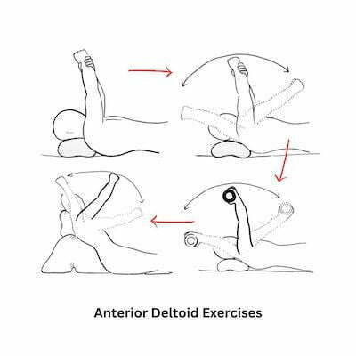
Σύνδρομο πρόσκρουσης ώμου ασκήσεις για τενοντίτιδα ώμου

Ασκήσεις - πόνος στον ώμο

Ασκήσεις για τενοντίτιδα ώμου

FAQs - Frequently Asked Questions

What is shoulder tendonitis?

Shoulder tendonitis is a common condition characterized by inflammation of the rotator cuff tendons, often resulting from repetitive overhead movements or structural impact.

How is shoulder tendonitis managed?

– Physiotherapy/home exercise program
– Cortisone or PRP injections
– Shoulder arthroscopy

Πόσος καιρός χρειάζεται για να περάσει η τενοντίτιδα ώμου?

Εξαρτάται από πολλούς παράγοντες, όπως την βαρύτητα της πάθησης και την προσήλωση του ασθενούς στη θεραπεία. Ήπιες περιπτώσεις μπορεί να βελτιωθούν σημαντικά σε 1-2 εβδομάδες, ενώ πιο βαριές τενοντίτιδες μπορεί να χρειαστούν εντατική θεραπεία και μερικούς μήνες.

Find us

Book an appointment with us today

Shoulder tendonitis | Diagnosis & treatment Read More »

Προβλήματα στο πόδι

9 foot problems you should know

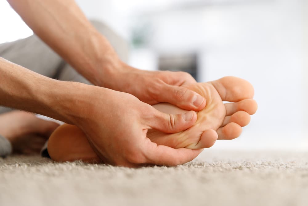
Our foot is a complex anatomical structure, consisting of 26 bones, 33 joints, 107 ligaments, 19 muscles and tendons. Numerically, the 52 bones of both legs make up 25% of the total 206 bones in our body. Research shows that on average we take 8,000 to 10,000 steps a day. 

Foot diseases are among the most common health problems. It is obvious that feet, like other parts of the body, need careful care. And yet, the health of our feet, this important part of the human body, is often overlooked. Here we will talk about some of the most important foot problems.

Plantar fasciitis

Plantar fasciitis is the most common cause of heel pain. Pain from plantar fasciitis is often most noticeable in the morning, during the first few steps after getting out of bed. The plantar fascia is a thick band of tissue on the sole of the foot. Repetitive loading can lead to microtears of the plantar aponeurosis, especially near the heel bone. This micro-tear leads to an inflammatory/healing response, which causes the pain.

Πελματιαία απονευρωσίτιδα

Risk factors for plantar fasciitis include: excessive standing, increased body weight, age, recent change in activity level, and stiff calf muscle.Some patients show a characteristic osteophyte (heel spur) on the X-ray, while others have a completely normal X-ray.

Plantar fasciitis can be treated non-surgically in the vast majority of patients. The main components of an effective conservative treatment program are: calf stretches with the knee extended, plantar fascia stretching, activity modification (to avoid hazardous activities), and wearing comfortable shoes. Shockwave therapy is also effective. In extreme cases, surgery may be required.

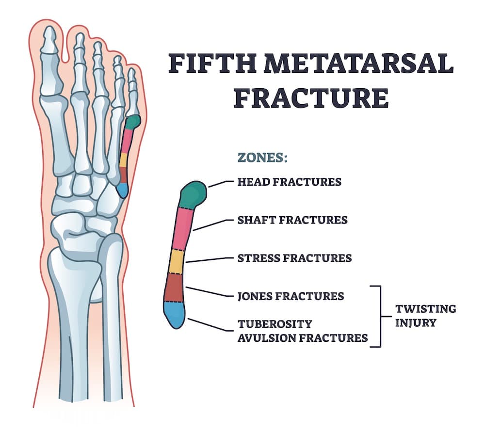
5th metatarsal fracture

The 5th metatarsal is the last bone on the outer surface of the foot. Fracture of the 5th metatarsal is very common in athletes, such as runners, football players, basketball players, who play sports that involve jumping and changing direction. The vast majority of these fractures are treated conservatively, with a boot. An exception are the so-called Jones fractures (zone 2), which are often difficult to heal, and may require surgery, especially in athletes.

shutterstock 2165496351
5th metatarsal fracture & Jones fracture

Posterior ankle impingement & Os Trigonum Syndrome

Posterior ankle impingement is characterized by pain behind the ankle joint. Symptoms worsen during plantar flexion of the foot. Posterior impingement is differentiated from insertional Achilles tendinitis and retrocalcaneal bursitis, as these conditions are more closely associated with the attachment of the Achilles tendon to the heel bone.

Os trigonum syndrome is also a result of an overuse injury to the posterior tibia caused by repetitive plantar flexion forces.It is mainly seen in ballet dancers and football players. The extra bone is usually visible on plain x-rays.

Treatment is usually non-surgical, with rest, activity modification, anti-inflammatories and occasionally a cortisone injection. If there is mechanical impingement, or a large extra bone behind the ankle (os trigonum), treatment may involve removing the extra bone.

Achilles tendinopathy & rupture

Achilles tendonitis is a chronic condition characterized by pain and often swelling in the Achilles tendon. The symptoms are due to swelling and inflammation of the tissue surrounding the Achilles tendon. There are 2 main categories, insertional and non-insertional tendinopathy. Treatment is mostly conservative, with rest, ice therapy, anti-inflammatories, an exercise program, and PRP. Surgery is indicated in resistant cases.

Achilles tendon ruptures  usually occur in athletic people in their 30s, 40s and 50s. The injury usually occurs after a sudden application of force resulting in the rupture (rupture) of the Achilles tendon.Achilles tendon rupture can be successfully treated conservatively or surgically. Recent studies suggest that nonsurgical and surgical treatment of Achilles tendon ruptures produce equivalent results.

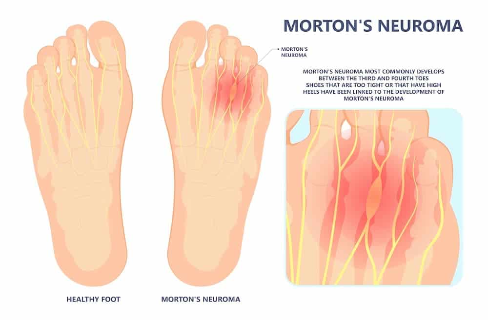
Morton's neuroma

Morton's neuroma is a painful condition that affects the sole of the foot, usually the area between the second and third toes. The sensation most patients report is that they are stepping on a pebble.

shutterstock 2023246769
Morton's neuroma

In Morton's neuroma, a thickening of the tissue develops around one of the nerves that innervate the toes. This can cause burning pain in the sole of the foot, and numbness or burning in the toes. Morton's neuroma appears to be the result of chronic irritation or repeated microtrauma to one of the nerves leading to the toes. If conservative treatment fails, a local injection of cortisone or anesthetic may be given. If this does not help, the next step is to surgically remove the neuroma. 

Bunions

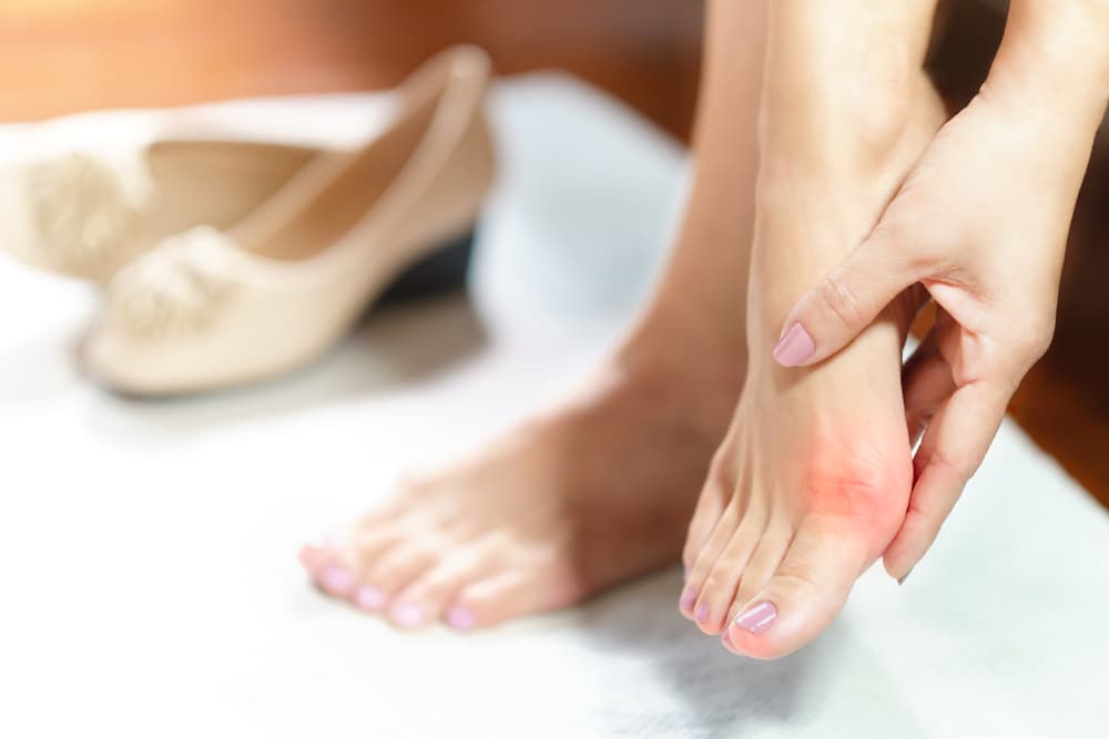
Hallux valgus, a condition also known as bunions, is one of the most common foot deformities. In this condition, the proximal phalanx of the big toe deviates outward resulting in the formation of an ossification (bone) on the medial side of the 1st metatarsophalangeal joint. A bunion is a relatively common condition. It occurs in about 23% of adults aged 18 to 65 and up to 36% of adults over 65.

Κότσι στο πόδι

The exact cause is not fully understood. It tends to happen more often in women and those who wear tight shoes or heels. Heredity also seems to play an important role. Based on x-rays of the feet, and depending on the angles formed between the bones, the condition is classified as mild, moderate and severe. Patients often report pain and difficulty walking. Initial treatment is conservative and aims to manage symptoms. In case of failure, surgical treatment is indicated. Usually, the surgery is done with a small incision, with an osteotomy of the first metatarsal (eg, Scarf, Chevron, Akin, etc.), accompanied by release of soft tissue and removal of the ossification.

Flatfeet

Flatfoot deformity is the condition in which there is a reduction in height or elimination of the foot arch. The incidence of the condition in the general population is high and can reach up to 30%.

Flatfoot is referred to as flexible, when the arch is absent during bearing but reappears when the foot is not bearing or standing on tiptoes. This form is benign – it causes no problems, or causes pain only with vigorous activity. Otherwise, we are talking about rigid flatfoot, which often coexists with serious diseases and causes pain during daily activities.

One of the most common causes of flat feet in adults is a dysfunction or insufficiency of the tibialis posterior tendon.

In most patients, a flexible flatfoot is asymptomatic and does not cause pain. In these cases, there is usually no cause for concern. Treatment of flat feet is indicated if pain is caused in the feet or soles, or if the condition affects the knees or lower back. Treatment may include special foot exercises, special arch support insoles, or splints.

In some cases, surgery can provide permanent relief. Common surgical procedures include flexor hallucis longus tendon transfer and calcaneal osteotomy for tibialis posterior insufficiency, or triple arthrodesis in advanced flat feet.

Arthritis

The most common forms of foot arthritis are:

  • Ankle arthritis: primary or post-traumatic.
  • Subtalar arthritis: often post-traumatic.
  • Hallux rigidus: arthritis of the 1st metatarsophalangeal joint.

The treatment here can be either conservative or surgical, depending on the area, severity, stage, etc.

Πόνος στο πόδι - κότσι

Toe deformities (hammertoe – claw toe – mallet toe)

Hammer toe is a common deformity of the toes. In hammertoes, the last phalanx of a toe is permanently bent toward the ground. As a result, a painful callus appears, both on the dorsal surface from the pressure of the shoe, and on the tip of the toe, from the pressure of the ground when walking. When the deformity is severe, the base of the toe bends and lifts away from the ground.Then, the angulation of the toe can become particularly acute, a condition called claw toe. Another similar condition is mallet toe. When conservative treatment fails, especially in painful and severe deformities, surgical treatment is required.

FAQs - Frequently Asked Questions

What's a bunion?

It is the deformity of the big toe, and it causes aesthetic and functional problems. It is painful and afflicts a large part of the female population. The big toe takes on an inclination towards the rest, while the first metatarsal projects in the opposite direction.

What's Os trigonum syndrome?

Os trigonum syndrome is also a result of an overuse injury to the posterior tibia caused by repetitive plantar flexion forces.It is mainly seen in ballet dancers and football players.

Find us

Book an appointment with us today

9 foot problems you should know Read More »

Πόνος στον ώμο

Understanding Shoulder Pain: Causes and Treatment Options

Shoulder pain is a common orthopaedic complaint that affects millions of people worldwide. The shoulder is a complex joint with a wide range of motion, making it susceptible to various injuries and conditions. In this blog article, we will explore the causes of shoulder pain and discuss some effective treatment options.

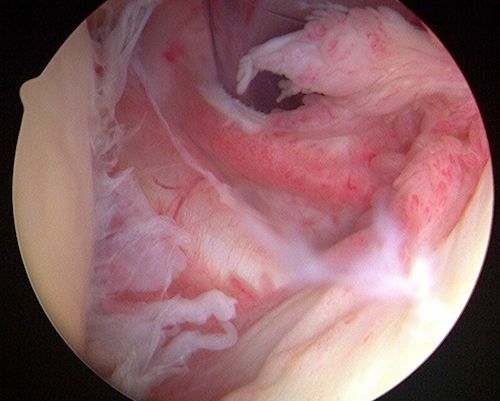
Παγωμένος ώμος - φλεγμονή του θυλάκου κατά την αρθροσκόπηση
Πόνος στον ώμο

Causes of Shoulder Pain

There many different conditions that can present with shoulder pain.

Rotator Cuff Injuries

The rotator cuff consists of a group of muscles and tendons that surround the shoulder joint, providing stability and facilitating movement. Overuse, repetitive motions, trauma, or aging can lead to rotator cuff tears or strains, causing pain, weakness, and limited range of motion.

Cuff tendonitis - Impingement syndrome

This occurs when the rotator cuff tendons become irritated or inflamed, often due to repetitive overhead activities. Shoulder impingement causes pain when raising the arm and can result in bursitis or tendonitis.

Frozen shoulder

Frozen shoulder is characterized by stiffness and pain in the shoulder joint. The exact cause is unknown, but it often develops after an injury or prolonged immobilization. People with diabetes or thyroid disorders are more prone to developing frozen shoulder.

Shoulder arthritis

Osteoarthritis and rheumatoid arthritis can affect the shoulder joint, leading to chronic pain, stiffness, and reduced mobility. Arthritis typically worsens over time and may require a combination of non-surgical and surgical interventions.

Shoulder instability

Shoulder instability occurs when the joint's structures, including ligaments and tendons, are unable to keep the humeral head (upper arm bone) securely within the shoulder socket. This can result in recurrent dislocations, causing pain and instability.

Calcific Tendonitis

Calcific tendonitis is caused by calcium deposits in the rotator cuff tendons, especially the supraspinatus. Exact cause remains unknown. It can cause intense pain, especially at night.

Shoulder fractures

Shoulder fractures may include proximal humerus fractures, clavicle fractures, scapula fractures, ACJ separations, etc.

Shoulder pain - Diagnosis

The doctor will begin the diagnostic approach with history taking and physical examination. The doctor will assess the range of motion and perform specific clinical tests for any possible condition. He will then perform an ultrasound of the area and order some further imaging tests depending on the possible underlying disease, such as X-rays and MRIs.

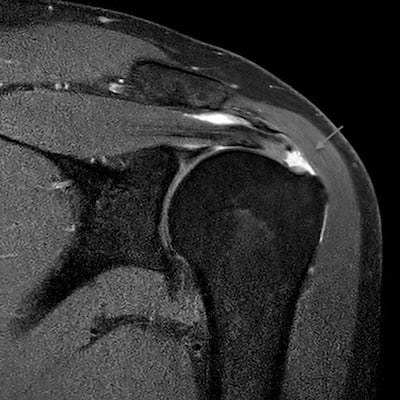
Παθήσεις ώμου - ρήξη τένοντα ώμου MRI
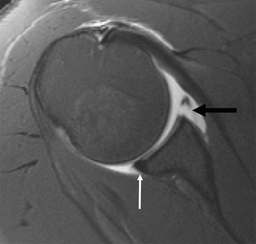
Εξάρθρημα ώμου - βλάβη Bankart se MRI

Shoulder pain - Treatment

Treatment options for shoulder pain may include:

1. Rest and Activity Modification: In many cases, shoulder pain can be managed with rest, avoiding activities that exacerbate the pain or strain the shoulder. Modifying your daily activities and using proper ergonomics can help alleviate symptoms and promote healing.

2. Physical Therapy: A tailored physical therapy program can help strengthen the muscles surrounding the shoulder joint, improve range of motion, and alleviate pain. Physical therapists use various techniques, including exercises, stretches, and manual therapy, to promote healing and restore function.

3. Medications: Non-steroidal anti-inflammatory drugs (NSAIDs) such as ibuprofen or naproxen can help reduce pain and inflammation in the shoulder joint. However, it's important to consult a healthcare professional before using any medication.

4. Corticosteroid Injections: In some cases, corticosteroid injections may be recommended to reduce inflammation and provide temporary pain relief. These injections are usually combined with other conservative treatments like physical therapy.

5. Surgical Intervention: If conservative treatments fail to alleviate shoulder pain or if the condition is severe, surgery may be necessary. Surgical options include arthroscopy, rotator cuff repair, shoulder replacement, or stabilization procedures, depending on the underlying cause of the pain.

Shoulder pain - prevention

Preventing shoulder pain involves maintaining good posture, practicing proper lifting techniques, and avoiding repetitive overhead activities. Regular exercise to strengthen the shoulder muscles and maintaining a healthy weight can also help reduce the risk of shoulder injuries.

Shoulder pain can significantly impact daily activities and quality of life. It is important to seek medical attention if shoulder pain persists or worsens despite conservative measures. An orthopaedic specialist can provide an accurate diagnosis and develop an appropriate treatment plan to address the underlying cause of the pain. 

Remember, each case of shoulder pain is unique, and treatment options may vary. Consulting with a healthcare professional is crucial for an individualized approach to managing shoulder pain effectively.

Underestimating the problem may lead to unnecessary delays, or lead to chronic issues that can become more difficult to eradicate. Please contact us to receive timely & personalised treatment for your pain.

FAQs - Frequently Asked Questions

What are the causes of shoulder pain?

– Impingement syndrome & Rotator cuff tears
– Dislocation/Instability
– Calcific tendonitis
– Fractures
– Arthritis
– Frozen shoulder

When should I see a doctor?

– Acute injury with severe pain
– Pain not mitigated by painkillers
– Night pain that disturbs sleep
– Pain that disrupts activities of daily living

Find us

Book an appointment with us today

Understanding Shoulder Pain: Causes and Treatment Options Read More »

Scroll to Top